The short answer: 3D when 2D is not enough
A panoramic X-ray gives a two-dimensional overview of both jaws and all the teeth. Cone beam computed tomography (CBCT) reconstructs a three-dimensional volume of a chosen area, which can be sliced in any direction. The rule is simple: 3D imaging is requested when a specific clinical question cannot be answered with a 2D examination, such as measuring bone width before an implant or locating a wisdom tooth in relation to the nerve.
Cone beam is therefore not an upgraded panoramic to be taken routinely. The two examinations answer different questions.
Everyday 2D X-rays and what they show
Several two-dimensional X-rays are used routinely in dental practice:
- The periapical X-ray: a small image of one to three teeth, from the crown to the root tip. It is very detailed and used especially for infections, root canal treatment and checking completed work.
- The bitewing X-ray: it shows the crowns of upper and lower teeth together and helps detect decay between teeth and early bone loss.
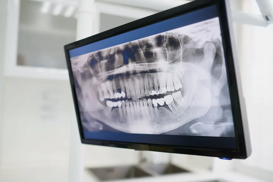
- The panoramic X-ray: a single image that unrolls both arches, impacted teeth, the maxillary sinuses, the mandibular nerve canal and the jaw joints.
The panoramic is valuable for a general assessment: a first visit, wisdom teeth, pre-orthodontic records, looking for a lesion. Its limits are well known: the image contains distortion and magnification that vary across areas, structures overlap and, above all, it does not show bone width from cheek to tongue. It is also less detailed than a periapical X-ray for examining a root closely.
What cone beam adds
In a cone beam scan, an X-ray source and a sensor rotate around the head in a single sweep. Software reconstructs a volume made of tiny cubes (voxels), which the clinician explores in axial, coronal and sagittal slices, or along the curve of the arch.
What it shows:
- bone height and width, with measurements that can be used to plan an implant;
- the exact position of a root relative to the mandibular canal or the sinus;
- the position of an impacted tooth, such as a canine stuck in the palate, and its relation to neighbouring roots;
- the extent of a cyst or lesion around a root;
- some complex root and canal anatomy in endodontics;
- the state of the bone walls after an extraction or an injury.
It has limits too. Metal items (crowns, posts, implants, root fillings) create artefacts, streaks that make the area around them hard to read. Soft tissue contrast is poor, so it is not an examination of the gums or muscles. And the fine detail of a small crack is not always visible, even in 3D.
When a 3D scan is genuinely useful
European guidelines on dental cone beam imaging, developed in particular through the SEDENTEXCT project, stress one point: every examination must be justified for the individual patient. The most common indications are:
- Implant dentistry: assessing bone volume, locating the nerve and the sinus, planning guided implant surgery or a bone graft.
- Lower wisdom teeth: when the panoramic shows signs of a close relationship with the nerve, 3D clarifies the real position and helps choose the technique or discuss an alternative.
- Impacted teeth: canines or other blocked teeth, before orthodontic traction or extraction.
- Complex endodontics: retreatment, a suspected extra canal, root resorption, or a persistent lesion not explained by 2D images.
- Cysts and bone lesions: measuring their extent and relations before surgery.
- Trauma: root or alveolar bone fractures when 2D images are inconclusive.
Conversely, cone beam is not indicated for routine caries screening, a simple annual check-up or a scan "just to see" without a specific question.
Dose and radiation protection: the ALARA principle
Every X-ray involves a small amount of ionising radiation. Medical radiation protection rests on three principles:
- Justification: the examination must provide useful information that may change the decision.
- Optimisation, summed up by the acronym ALARA (as low as reasonably achievable): obtaining the image needed with the lowest reasonable dose. The term ALADA is also used, to match the dose to the diagnostic goal.
- Limitation: choosing the right examination without taking unnecessary images.
In practice, the order of magnitude is as follows: digital intraoral X-rays deliver a very low dose, a panoramic a low dose, and a cone beam a higher dose than a panoramic, which varies with the device, field of view and resolution, while generally remaining lower than a conventional medical CT scan. The main lever is the field of view: a small field centred on two or three teeth exposes far less than a volume covering both jaws.
Children and teenagers are more sensitive to radiation, so justification is even stricter for them. If you are pregnant, always say so: the examination is postponed if it can wait, or carried out with appropriate precautions if it is needed. To learn more, read our article are dental X-rays dangerous?
What the examination is like and questions to ask
A panoramic is taken standing or sitting, with your chin on a rest and your teeth biting a small block: the machine rotates around your head for a few seconds. A cone beam scan is very similar. You need to remove jewellery, piercings, glasses and removable dentures, and stay completely still, because movement can blur the image and mean a retake.
Reading a cone beam scan is not limited to the area requested: the whole volume should be reviewed by a trained professional, as it can show unexpected findings in the sinuses or bones.
Useful questions to ask:
- What specific question is this scan meant to answer?
- Would a 2D X-ray not be enough?
- What field of view is planned, a limited area or both jaws?
- Can I have the images and the report for my records?
Keeping your previous X-rays can sometimes avoid repeating an examination.
Imaging at German Dental Clinic in Agdal
Dr Fenjero Walid's practice has digital radiography and a panoramic X-ray unit, used for the initial assessment and to monitor treatment. When your situation justifies three-dimensional imaging, for example before a dental implant or the removal of a wisdom tooth close to the nerve, the dentist explains why, prescribes the scan and goes through it with you before proposing a treatment plan.
If you live in Rabat and are unsure about an examination you have been asked to have, you can book an appointment and bring your existing X-rays.
Frequently asked questions
Why does my dentist want a cone beam scan when I already have a panoramic?
Because a panoramic and a cone beam scan answer different questions. The panoramic gives a 2D overview, without measuring bone width or precisely locating a root in relation to the nerve. When the decision depends on that information, for example before an implant, 3D becomes useful. Your dentist should be able to explain what they are looking for.
Is a cone beam scan dangerous?
A cone beam scan delivers a higher dose than a panoramic, but generally lower than a medical CT scan, especially with a small field of view. The risk is low when the examination is justified and optimised. That is precisely why it is not done routinely, only when it provides information that matters for your treatment.
Can I have a dental X-ray during pregnancy?
Always mention a pregnancy or a possible one. Dental X-rays target the mouth, far from the abdomen, but extra caution applies: a non-urgent examination is usually postponed until after the birth. If an X-ray is needed to treat pain or an infection, it is taken with suitable protection and with as few images as possible.
Is a cone beam scan needed before every implant?
For most implant projects, 3D imaging helps measure the bone and locate the structures to avoid, and it is essential for guided surgery. In some simple cases, the dentist may decide that it would not add decisive information. The decision depends on the area, the visible bone volume and the type of procedure planned.
Should I keep my old X-rays?
Yes, they are very useful. Previous X-rays make it possible to compare how a lesion, bone loss or a treatment has changed over time, and can sometimes avoid a repeat examination. Digital images can be kept on a USB stick or by e-mail. Bring them to your consultation, especially if you are changing dentist.
The information on this page is general. Only a consultation allows a diagnosis and a suitable treatment plan.