What is an impacted canine?
A canine is called impacted when it stays trapped in the jawbone beyond the usual age of eruption, even though its root is already well formed. It is most often the upper canine, frequently lying on the palate side. Treatment depends mainly on age and on the tooth's position: in a child, early detection sometimes allows the tooth to come through on its own; later, surgical exposure is usually combined with orthodontic traction, and extraction is kept for cases where the tooth cannot be guided into place.
The canine is no ordinary tooth. It has the longest root in the mouth, shapes the corner of the smile and guides the jaw in side-to-side movements, which protects the back teeth. Losing it or leaving it aside is never a trivial matter.
Several factors explain why it gets stuck:
- its eruption path is the longest of all teeth, starting high up near the nose;
- lack of space in the dental arch;
- a missing or small lateral incisor, which no longer acts as a guide;
- a baby canine whose root does not resorb normally;
- a family tendency, which is often found;
- more rarely, an obstacle such as a cyst or an extra tooth.
When to worry: the warning signs
The upper adult canine usually erupts between 11 and 13 years of age, with wide variation between children. A delay on its own is therefore not a disease. Some signs, however, call for an examination:
- the baby canine stays firmly in place while the other baby teeth in that area have fallen out;
- a canine has come through on one side but not the other for several months;
- around the age of 10, the dentist cannot feel the bulge of the canine under the gum above the baby tooth;
- the neighbouring lateral incisor tilts, drifts apart or becomes mobile;
- a hard bump appears on the palate.
An impacted canine is usually painless. It is therefore often found by chance, at a check-up or on a panoramic X-ray taken for another reason. Adults sometimes only learn about it many years later, when the baby canine starts to wobble.
Regular check-ups between 9 and 11 are genuinely useful here: this is the age when simple measures still work. For the general timetable of tooth loss, see our article on baby teeth.
Diagnosis: clinical examination and imaging
Diagnosis starts with the examination: feeling the gum and palate, looking at the position of the incisors, the space available and how loose the baby canine is.
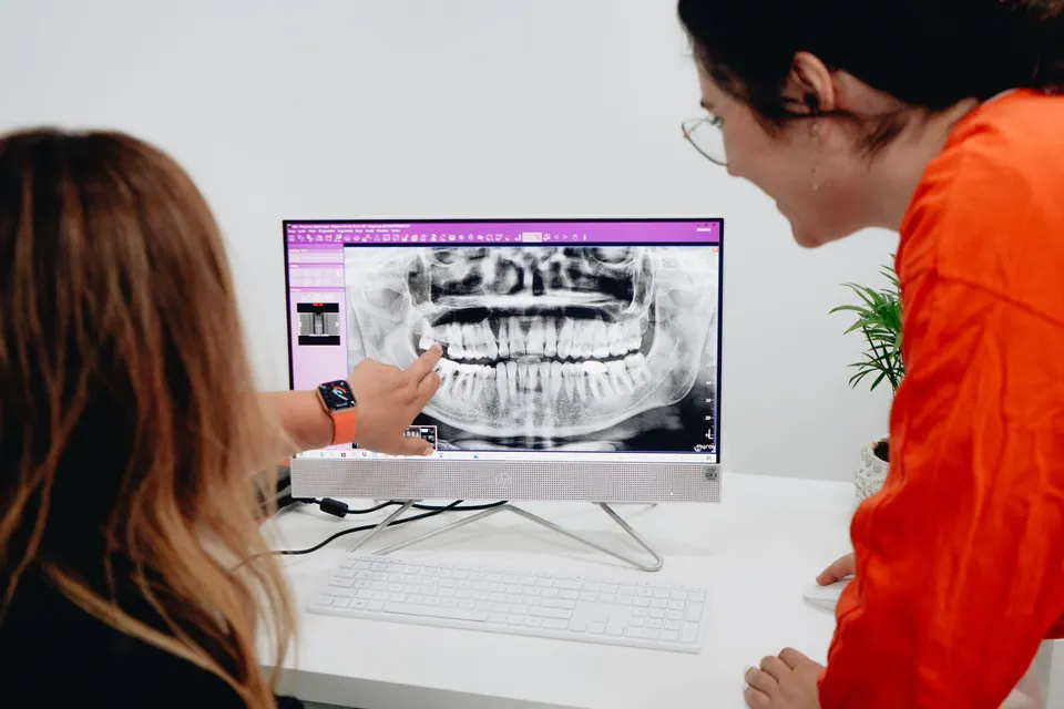
A panoramic X-ray confirms that the canine is there and gives a first idea of its position: its angle, its height and whether it overlaps the root of the neighbouring incisor. Additional images taken from different angles can help tell whether the tooth lies on the palate side or the lip side.
When treatment is being considered, three-dimensional imaging such as cone beam CT is often useful. It locates the crown precisely and, above all, shows whether the canine has started to resorb the roots of the neighbouring incisors, a silent complication that changes treatment priorities. Your dentist will tell you whether this scan is justified in your case; our article on 3D cone beam imaging explains what it adds and the principle of the lowest reasonable dose.
By the end of the assessment, several questions have an answer:
- can the tooth still come through on its own?
- does space need to be created in the arch?
- is traction realistic given its position?
- are the neighbouring teeth at risk?
In children: intercepting early
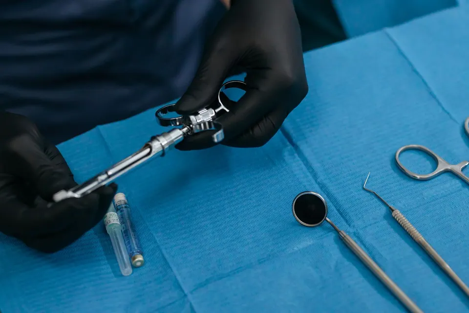
When the canine is spotted early, around 10 to 12 years of age, and is not too far off course, a simple measure may be enough: removing the baby canine. Freed from this obstacle, the adult canine sometimes changes direction and eventually erupts by itself. The procedure is quick, done under local anaesthetic and well tolerated by most children.
This approach is not automatic. Its chances depend on the canine's angle, its height and the child's age: the more horizontal the tooth and the closer the child is to adolescence, the less likely a spontaneous correction becomes. The orthodontist may also suggest creating space in the arch, for example with an expansion appliance, to make eruption easier.
A follow-up X-ray is then planned after several months to check that the canine is moving in the right direction. If it has not moved, the next step is taken without losing more time.
This window really matters: a canine guided early often avoids longer surgery and traction in the teenage years. Parents who notice a baby canine that is still very firm at 11 or 12 are right to ask for an opinion.
Surgical exposure and orthodontic traction, step by step
In teenagers, and sometimes in adults, the standard treatment is to reach the canine and pull it gently into place. It relies on close teamwork between the dental surgeon who operates and the orthodontist who manages the traction.
- Preparing the space: the orthodontist first fits an appliance, usually fixed braces, to open enough room and provide solid anchorage.
- Exposing the crown: under local anaesthetic, the gum is opened and the bone covering the tip of the canine is carefully removed, without touching its root.
- Bonding an attachment: a small button with a fine chain is bonded to the crown.
- Closing: in the closed technique, the gum is put back and only the chain shows; in the open technique, used mainly for canines on the lip side, the gum is repositioned to keep a firm band of gum around the tooth.
- Pulling slowly: light forces, adjusted at each visit, guide the tooth over several months.
- Aligning and stabilising: once the canine is in the arch, alignment is completed and a retainer holds the result.
Recovery after surgery is usually straightforward: moderate swelling and discomfort for a few days. Complications do exist: the attachment coming off and needing to be rebonded, gum recession, or the discovery of ankylosis, meaning the tooth is fused to the bone and does not respond to traction.
Clear aligners can play a part in some treatment plans, particularly for final alignment, but the traction phase usually requires fixed components: the orthodontist decides.
Extraction or keeping the baby canine: the other options
Bringing the canine down is not always possible or desirable. Removing it is considered in particular when:
- it is ankylosed or has not responded to traction;
- its position is too unfavourable, lying horizontally or very high;
- it is resorbing the roots of the neighbouring incisors;
- a cyst has formed around its crown;
- the adult does not want lengthy orthodontic treatment.
This is a more delicate procedure than a simple tooth extraction, often through the palate, with a few days of possible swelling.
An impacted canine causing no harm can also be left in place and monitored with X-rays at long intervals, especially in adults, provided it threatens neither the neighbouring roots nor the bone.
As for the baby canine that stays, it can sometimes serve for many years in an adult. Being smaller and more yellow, it can be reshaped with composite resin to blend into the smile. Its root often resorbs in the end, though, so it is wise to plan its replacement before it falls out. Depending on the situation, options then include an implant once growth is complete, a bridge, or closing the space orthodontically, with the premolar reshaped to look like a canine.
Having an impacted canine assessed in Agdal
Whether you are the parent of a 10-year-old whose baby canine will not budge, or an adult who has just found an impacted canine on an X-ray, the question is the same: what can be done now, and what is the risk of waiting?
At German Dental Clinic in Agdal, the assessment combines a clinical examination with digital panoramic radiography, supplemented by prescribed 3D imaging when needed. The surgical part, exposure or extraction, falls within oral surgery; traction is carried out by an orthodontist with whom the plan is coordinated, and aligners are discussed when they are suitable.
For an opinion in Rabat, bring any recent X-rays you have and book an appointment.
Frequently asked questions
Does an impacted canine hurt?
Usually not. An impacted canine can stay in the bone for years without any symptoms, which is why it is often found by chance on an X-ray. Pain, swelling or a loose incisor can, however, signal a complication such as resorption of the neighbouring roots or a cyst, and call for a prompt examination.
Can an impacted canine be brought down in adulthood?
It is possible in some cases, as there is no strict age limit. Treatment is less predictable than in adolescence, though: traction is often slower and the risk of ankylosis, a tooth fused to the bone, increases with age. The X-ray assessment and the orthodontist's opinion show whether an attempt is reasonable.
Is surgical exposure painful?
The procedure is done under local anaesthetic and should not hurt. In the following days, discomfort and moderate swelling are common and are usually well relieved by the prescribed medication. Soft food, gentle brushing around the area and following the instructions help it heal without trouble.
Are clear aligners enough to treat an impacted canine?
Rarely on their own. Pulling an impacted canine into place usually requires fixed components bonded to the teeth and managed by an orthodontist. Aligners can, however, be used in certain phases, for example to create space or for finishing, depending on the treatment plan drawn up for you.
Should an impacted canine that is not treated be removed?
Not necessarily. If it does not threaten the neighbouring roots, shows no cyst and the patient does not want treatment, it can be left in place and checked with regular X-rays. Extraction is suggested when it causes damage or gets in the way of another plan, such as placing an implant.
The information on this page is general. Only a consultation allows a diagnosis and a suitable treatment plan.
A question about your own situation?
Dr Fenjero Walid sees patients in Agdal, Rabat, for an assessment and answers that fit your case.