What is a dental cyst?
A dental cyst is a cavity in the jawbone, lined with a thin membrane and filled with fluid, that most often develops at the tip of the root of a tooth whose nerve has died and become infected. It grows slowly, is often painless and is frequently found by chance on an X-ray. It does not go away on its own while its cause remains: treatment means removing the infection, through treatment of the tooth, minor surgery or sometimes an extraction.
The most common form is the radicular cyst, also called a periapical cyst. It almost always starts the same way: deep decay, a fracture or an old knock has allowed bacteria to reach the pulp, the living tissue at the centre of the tooth. The pulp dies, bacteria colonise the canals, and the body reacts at the root tip by forming an area of chronic inflammation that gradually destroys the surrounding bone.
The word cyst often sounds alarming. It helps to know that the vast majority of jaw cysts are benign: they are not malignant tumours. They still need attention, though, because they can enlarge, weaken the bone and eventually become infected.
Granuloma or cyst: what is the difference?
On an X-ray, the lesion at the root tip shows up as a dark area, where bone has been replaced by soft tissue. That tissue can be of two kinds:
- a granuloma: a mass of chronic inflammatory tissue with no cavity, representing the bone's defensive reaction to infection coming from the tooth;
- a radicular cyst: a true sac, lined with a layer of cells (an epithelium) and containing fluid. It often develops from a long-standing granuloma, from cells naturally present around the root.
An X-ray alone cannot tell them apart with certainty. A large lesion with very sharp borders points more towards a cyst, but only microscopic examination of the removed tissue, known as histopathology, confirms the diagnosis. That is why dentists often speak, more cautiously, of a periapical lesion or apical periodontitis.
In practice, the distinction changes the decision less than you might think: a granuloma and a radicular cyst share the same cause, an infected tooth, and the first stage of treatment is usually the same. It is how the lesion responds to that treatment that guides what happens next.
Other cysts of the jaws
Not every cyst in the mouth comes from an infected tooth. Depending on where it sits and how it looks, the dentist may consider others:
- A follicular (dentigerous) cyst surrounds the crown of a tooth that has stayed buried in the bone, very often a lower wisdom tooth or a canine. It is frequently discovered during an assessment before extraction.
- A residual cyst remains in the bone after an infected tooth has been extracted, when the lesion was not completely removed.
- Lateral and paradental cysts form on the side of a root, sometimes linked to a gum pocket.
- The odontogenic keratocyst is a particular type that can spread within the bone and is known to recur. It needs specific management and monitoring, often with an oral and maxillofacial surgeon.
Some other findings can look like a cyst without being one. That is why a lesion that does not match a diseased tooth, or does not heal as expected, should be analysed and sometimes referred to a specialist.
Symptoms: often none, sometimes signs to watch for
A dental cyst usually progresses without pain for a long time. That is why it is so often found on a panoramic X-ray taken for another reason: a check-up, planning for an implant or orthodontics, a wisdom tooth.
When signs do appear, they may include:
- a tooth that has changed colour, greyish or darker than its neighbours, a common sign of a dead nerve;
- discomfort when biting, or a feeling that the tooth is higher than the others;
- a small spot on the gum (a sinus tract or fistula) that discharges pus from time to time and then disappears;
- a firm, fairly painless swelling of the gum or palate, as the bone may bulge slightly when the lesion grows;
- neighbouring teeth that shift or become loose;
- a painful, rapid swelling if the cyst becomes infected, which then resembles an abscess.
One sign calls for a prompt appointment: numbness of the lip, chin or cheek. It can reflect pressure on a nerve and should always be investigated.
Diagnosis: tests, X-rays and sometimes a biopsy
Faced with a suspicious image, the dentist first tries to establish which tooth is involved and whether it is still alive.
- Pulp sensitivity tests (cold, sometimes an electric test): a tooth that does not respond suggests a dead pulp, and therefore a lesion of dental origin. A tooth that responds normally means another explanation must be looked for.
- Percussion and palpation tests, probing of the gum and looking for a sinus tract.
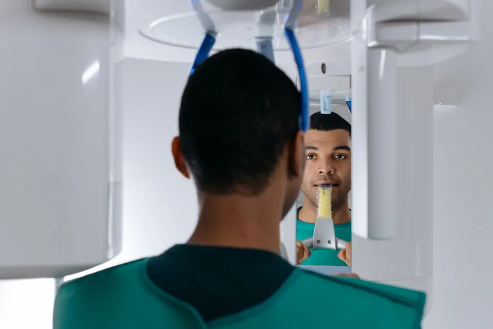
- A periapical X-ray, a small image focused on the tooth, and a panoramic X-ray, which gives an overview of both jaws and the size of the lesion.
- Three-dimensional imaging (cone beam CT), when indicated, shows the extent of the lesion and its relationship to the nerve of the lower jaw or the sinus. Your dentist will tell you whether this scan is useful in your case, following the principle of a dose as low as reasonably achievable.
When the lesion is removed surgically, the tissue is normally sent to a laboratory for histopathological examination. This is what establishes the exact nature of the cyst with certainty and rules out rarer lesions.
Treatment: treat the tooth, operate or extract
Treatment aims to remove the cause, then let the bone rebuild itself. The choice depends on the tooth, the size of the lesion and how it evolves:
- Root canal treatment: for a tooth that has never been treated, cleaning, disinfecting and filling the canals cuts the lesion off from its source of bacteria. Many periapical lesions, including some cystic ones, then shrink on their own. Our root canal treatment page describes the procedure.
- Root canal retreatment: if the tooth has already been treated and the lesion persists or appears, redoing the canal treatment is often the first option. The causes of failure and the steps are covered in our article on root canal retreatment.
- Apical surgery with enucleation: when the lesion does not heal after proper treatment, is large, or cannot be reached through the tooth, the dentist opens the gum, removes the whole sac, treats the root tip and sends the tissue for analysis.
- Extraction with curettage: if the tooth cannot be saved (fractured root, too much destruction, insufficient support), it is removed and the lesion carefully cleaned out. Replacing the tooth is then planned. See our tooth extraction page.
For very large cysts, decompression or marsupialisation techniques, which gradually reduce the volume before a second procedure, may be offered in a specialist setting. Antibiotics, for their part, do not treat a cyst: they only help with an acute infection that is spreading.
X-ray follow-up and assessment in Agdal
Bone healing cannot be seen within a few days. After treatment, a follow-up X-ray is generally scheduled after several months and repeated if needed, to check that the dark area is shrinking and bone is re-forming. A lesion that gets smaller is a good sign, even if the image does not return to completely normal straight away; a lesion that grows or stays the same means the next step needs to be reconsidered.
In the meantime, a few habits help: protect the root-treated tooth with a suitable restoration, keep up careful hygiene and report any swelling, sinus tract or numbness.
At German Dental Clinic in Agdal, digital panoramic radiography and targeted X-rays make it possible to analyse an incidental finding and explain the options clearly. If you have been told you have a dental cyst and live in or around Rabat, you can book an appointment for an opinion based on your X-rays.
Frequently asked questions
Is a dental cyst dangerous?
In the vast majority of cases it is a benign lesion linked to an infected tooth. Without pain or swelling it is not an emergency, but it should not be ignored: as it grows it destroys bone and can shift neighbouring teeth or suddenly become infected. Analysis of the removed tissue also rules out rarer lesions that need specific management.
Can a dental cyst go away on its own?
Not while the cause is still there: an infected tooth keeps feeding the lesion. Once the canals have been properly treated, however, many periapical lesions gradually shrink and the bone rebuilds itself without surgery. A follow-up X-ray, taken several months after treatment, is what confirms this progress.
Does the tooth have to be extracted?
No. When the tooth can be saved, the priority is to keep it with root canal treatment or retreatment, possibly followed by apical surgery. Extraction is considered when the root is fractured, the tooth is too badly damaged or its bone support is insufficient. The decision is made after a clinical and radiographic examination.
Are antibiotics enough to treat a dental cyst?
No. Antibiotics do not reach the inside of a tooth whose pulp has died, nor the contents of a cyst. They can calm a spreading infection with fever or significant swelling, but the lesion comes back if the tooth is not treated. They support dental treatment; they do not replace it.
Can an implant be placed after a cyst has been removed?
Often yes, but not always straight away. The bone must heal first, then the remaining volume is assessed with imaging. If the cavity left by the cyst was large, bone reconstruction may be proposed before or during implant placement. The timing depends on the size of the lesion and the quality of healing.
The information on this page is general. Only a consultation allows a diagnosis and a suitable treatment plan.